
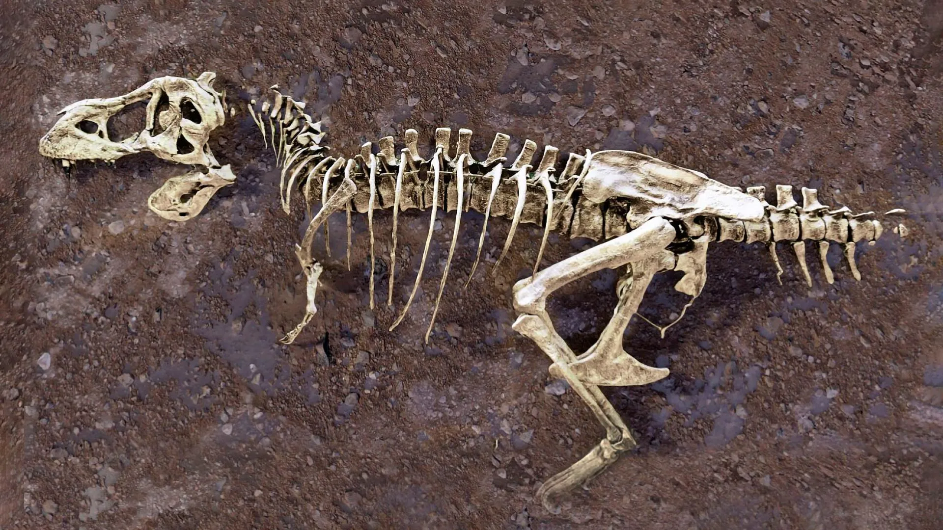
Scotty, the largest Tyrannosaurus rex skeleton ever discovered, has been dead for roughly 66 million years, which is normally plenty of time for soft tissue to vanish without a trace. But a fractured rib from the celebrated carnivore has handed researchers an almost unheard-of prize: fossilized blood vessels preserved inside the bone, evidence of an injury that was still healing when Scotty died.
Scientists examined the fossil using neutron imaging at the Department of Energy's Oak Ridge National Laboratory (ORNL), building detailed 3D images of the rib's interior without damaging the preserved soft tissue. "It's like winning the lottery," said Mauricio Barbi, professor of physics at the University of Regina (U of R) in Saskatchewan, Canada. "Scotty's rib contains a vast network of mineralized blood vessels that has never before been observed in a fossil."
Soft tissues like blood vessels typically disappear during decomposition long before fossilization finishes, making Scotty's rib a rare exception. After the rib broke, iron-rich blood flooded the injured area and new blood vessels formed as part of the healing process. Scotty died before the fracture fully healed, then ended up in a salty marsh where conditions slowed decomposition and helped preserve the fragile vessel network. "Every fossil is a tiny snapshot of the past," said Jerit Mitchell, a U of R doctoral candidate in physics who leads the project under Barbi's direction.
Scotty's remains were discovered by teams from the Royal Saskatchewan Museum in Saskatchewan's Frenchman River Valley, one of North America's richest dinosaur fossil sites. Rocks there preserve a key record of dinosaur life shortly before the mass extinction that ended the age of nonavian dinosaurs. Researchers are also studying fossilized amber, dinosaur scales and bones from other dinosaurs. "By piecing the clues together, we understand the past and how things could evolve in the future," said Marcella Berg, a U of R assistant professor of physics and former ORNL postdoctoral researcher.
Neutron and X-ray imaging offer complementary ways to peer inside materials. Neutrons excel at detecting light elements, especially hydrogen, while X-rays are highly effective at revealing heavier elements. The contrast is somewhat like the difference between an MRI, which emphasizes soft tissues such as muscle, and an X-ray, which is especially useful for viewing dense structures such as bone. Researchers pick different neutron and X-ray techniques depending on what material they want to investigate.
The work traces back to 2020, when Mitchell, then a U of R undergraduate, detected evidence of blood vessels inside Scotty's rib. At the Canadian Light Source, he first used micro-CT scanning, a noninvasive X-ray imaging method. The scans confirmed fossilized soft tissue in cut sections of the rib. As the investigation expanded, researchers combined other X-ray methods, including synchrotron radiation at the Canadian Light Source, with microscopy, allowing them to study both the healing injury and preserved fossil tissues at the cellular level. Once X-ray imaging exposed evidence of the fossilized blood vessels, the researchers turned to neutrons for additional clues.
In April 2026, the team used the Multimodal Advanced Radiography Station (MARS) instrument at ORNL's High Flux Isotope Reactor (HFIR) and the Virtual Environment for Neutron Sciences (VENUS) instrument at DOE's Spallation Neutron Source (SNS) at ORNL. Neutron imaging confirmed earlier observations, let the team examine large bones including Scotty's rib without damaging them, and provided extra image contrast that complemented the other methods. "Neutrons not only corroborated what we found with synchrotron radiation techniques that led to the discovery of blood vessels in Scotty's rib, but they also proved to be a highly valuable addition to our current studies in search of soft tissue preservation in fossils," Berg said. "This gives us an incredible amount of detail to better understand these properties without affecting the samples."
MARS generates cold neutrons that are particularly effective at highlighting signatures associated with soft tissue, areas rich in hydrogen and subtle differences within a specimen. Researchers used it to produce high-resolution images of smaller bones, amber and fossilized scales. VENUS, by contrast, produces high-energy neutrons capable of penetrating deeper into large objects and generating detailed 3D images, which the team used to examine larger bones including Scotty's rib.
As neutrons pass through a specimen, they interact with atoms throughout the material and are especially sensitive to hydrogen atoms. Those interactions generate information that can be converted into images. Because neutrons behave differently from X-rays, they can expose features that may be difficult or impossible to detect with other imaging technologies. "People often think of neutrons as tools for studying batteries or advanced materials, but they're just as innovative for answering questions about ancient life," said Hassina Bilheux, lead instrument scientist for VENUS.
Researchers will continue studying the data collected at VENUS and MARS while extending the approach to additional fossils, and they plan to compare patterns of injury and healing among different species. By combining neutron imaging with X-ray techniques, the scientists hope to investigate pathologies preserved in fossils and examine how those ancient conditions compare with differences found in modern species. "There are more fossils than you think sitting in collections, hiding secrets from millions of years ago," Mitchell said. "Putting them in a synchrotron or neutron source allows us to make new discoveries about ancient life like never before."
Materials provided by Oak Ridge National Laboratory. Note: Content may be edited for style and length.