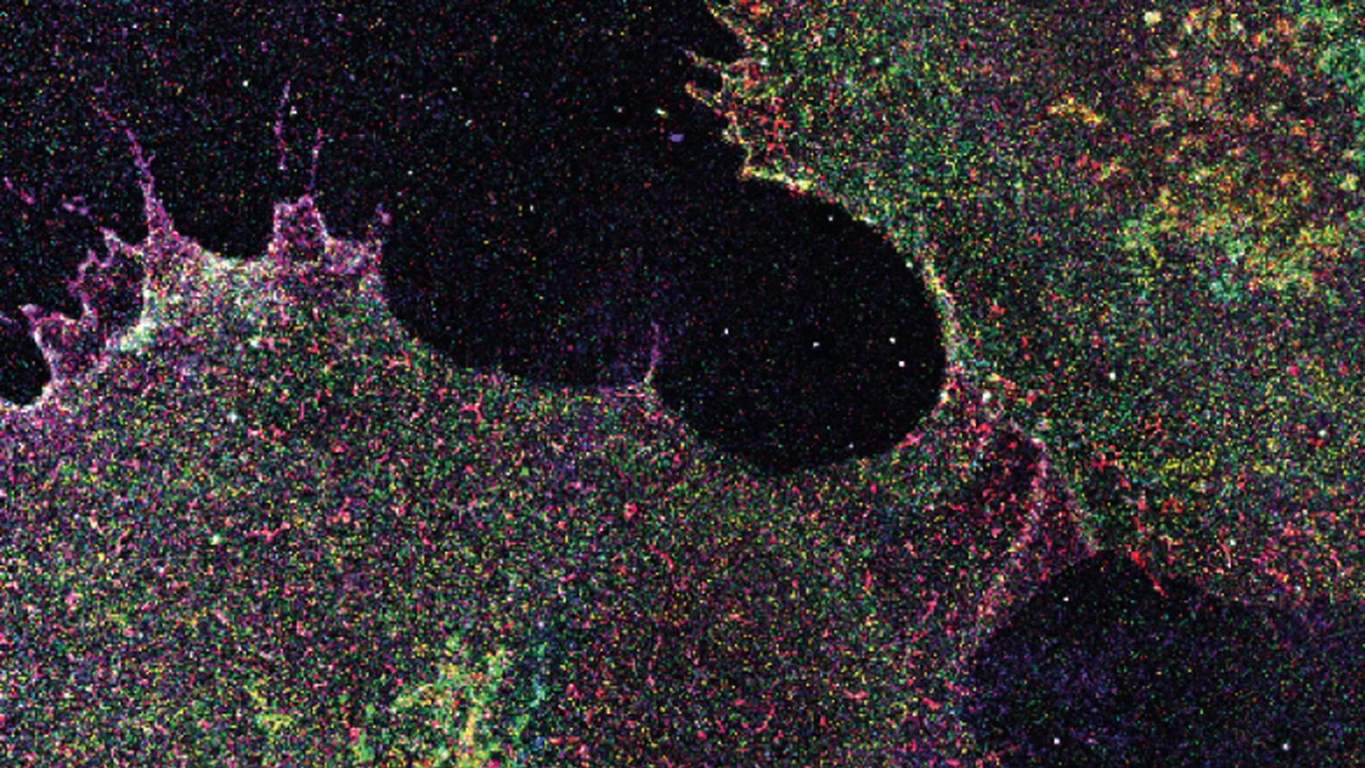
Every human cell wears a thin layer of sugar called the glycocalyx, which is less about candy and more about a protective outer shell that’s constantly shifting and reorganizing. Researchers at the Max Planck Institute for the Science of Light (MPL) have now created detailed maps of these sugar structures using advanced high resolution microscopy, and their findings, published in Nature Nanotechnology, suggest that changes in the arrangement of these sugars could one day help doctors detect diseases such as cancer.
The team, led by Prof. Leonhard Möckl in the “Physical Glycosciences” research group, developed a technique called “Glycan Atlasing.” Using cutting edge super resolution microscopy, they mapped the glycocalyx at the level of individual sugar molecules across many types of cells, including cell culture lines, primary human blood cells, and tissue samples. The resulting maps showed that the glycocalyx changes its molecular arrangement depending on the condition of the cell.
For example, immune cells displayed different sugar patterns after being stimulated, similar to what happens during an immune response. According to the researchers, this provides the first direct evidence that the glycocalyx functions almost like a display screen, showing information about a cell’s internal state on its outer surface. The team found that these nanoscale sugar patterns could reliably distinguish between different cellular states, allowing them to identify separate stages of cancer development, tell the difference between activated and inactive immune cells, and distinguish cancerous regions from healthy regions in human breast tissue.
“The results provide a promising foundation for the development of future diagnostic methods, as Glycan Atlasing delivers reliable results even in complex samples,” explains Möckl. The researchers now plan to expand the method by analyzing additional target structures, automating more of the process, and studying much larger numbers of samples so the technique can eventually be adapted for routine medical use. “In large-scale studies, we want to investigate which surface patterns are associated with specific disease courses or therapeutic responses and how cell states can be detected early and objectively via the surface,” Möckl says.
The Good Times
News in your inbox.
One sardonic roundup, delivered on your schedule. Free. Unsubscribe whenever your tolerance for wit runs out.
Already subscribed but we never reach your inbox? Check your spam folder and hit 'Not spam' (or 'Remove from spam') to bust us out of junk-mail purgatory. You'll be helping everyone else too.
Don't open any of our emails for a month and you'll be automatically removed from the mailing list.
Rewrite Article
Select parts to regenerate with a fresh AI pass. Translations will be updated automatically.
Generate AI Image
Creates a sardonic version of the article image using OpenAI.